microscope parts and functions pdf
Summary
Discover the essential microscope parts and their functions in our detailed PDF guide. Perfect for students, professionals, and enthusiasts. Download now!

A microscope is an essential scientific tool for exploring microscopic worlds. Understanding its parts and functions is crucial for effective use in various fields like biology, medicine, and research.
1.1 Overview of the Microscope and Its Importance

A microscope is a fundamental scientific instrument used to magnify small objects or samples, revealing details invisible to the naked eye. Its importance spans various fields, including biology, medicine, and materials science, enabling researchers to study microorganisms, cellular structures, and material compositions. The microscope has revolutionized scientific discovery, contributing to breakthroughs in disease diagnosis, drug development, and materials engineering. Understanding its structure and functions is essential for maximizing its potential in both educational and professional settings. Proper use ensures accurate observations and advancements in numerous disciplines.

1.2 Brief History of Microscope Development
The microscope has a rich history dating back to the late 16th century when the first compound microscope was invented by Zacharias Janssen in 1590. Over the centuries, advancements in optics and design led to improved models, with significant contributions in the 17th and 18th centuries. The 20th century saw the introduction of electron microscopes, which allowed for even greater magnification. This evolution has enabled scientists to study microorganisms, cells, and materials in unprecedented detail, revolutionizing fields like biology, medicine, and materials science.

Optical Components of the Microscope
The microscope’s optical system includes the eyepiece, objective lenses, and condenser. These components work together to magnify and focus light, enabling clear specimen observation at various resolutions.
2.1 Eyepiece Lens (Ocular Lens)
The eyepiece lens, or ocular lens, is a critical optical component that magnifies the image produced by the objective lens. Typically, it has a magnification power of 10×, though higher powers like 15× or 20× are available. It works in conjunction with the objective lens to provide the total magnification of the specimen. The eyepiece is designed to focus the light from the objective, creating a virtual image for the user to observe. Proper cleaning with a soft cloth is essential to maintain clarity and avoid scratches. Regular maintenance ensures optimal performance.

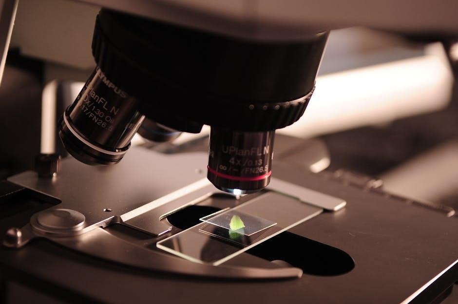
2.2 Objective Lenses
Objective lenses are crucial for capturing and focusing light from the specimen, delivering it to the eyepiece. They are mounted on a rotating nosepiece, allowing easy switching between different magnifications. Common magnifications include 4×, 10×, 40×, and 100× oil immersion. Each lens is designed for specific viewing needs, with higher magnification lenses requiring immersion oil to enhance resolution. Proper alignment and focus ensure sharp, clear images. Regular cleaning with specialized solvents prevents contamination and maintains optical clarity, ensuring accurate observations in scientific and educational settings.
2.3 Condenser Lens
The condenser lens focuses light from the illuminator onto the specimen, enhancing image clarity and resolution. Positioned beneath the stage, it ensures even light distribution across the specimen. The condenser adjusts light intensity and alignment, optimizing visibility. Its aperture diaphragm regulates light entry, balancing brightness and contrast. Proper condenser alignment is vital for sharp images, especially at higher magnifications. Regular maintenance, like cleaning, prevents light obstruction. This component is essential for achieving clear, detailed observations in microscopy, making it a key part of the optical system.

Structural Components of the Microscope
The microscope’s structural components form its backbone, supporting the optical system and specimen handling. These parts ensure stability, alignment, and precise control during observations, making them indispensable for functionality.
3.1 The Stage and Its Functions
The stage is a flat platform that holds the specimen in place under the microscope. It is typically equipped with clips or a mechanical stage that allows for precise movement of the specimen along the x and y axes. This ensures that different parts of the sample can be easily viewed without disturbing the setup. The stage’s durability and stability are crucial for maintaining focus and preventing vibrations during observation. It is an essential component for accurate specimen examination.
3.2 The Body Tube and Its Role
The body tube connects the eyepiece and objective lenses, housing the optical components. It maintains the correct distance between the lenses for proper image formation. The tube’s rigidity ensures alignment and prevents optical distortions. In some microscopes, the body tube is inclined for ergonomic viewing. Its durability is crucial for maintaining the microscope’s performance and longevity. The body tube plays a vital role in transferring light from the objective lens to the eyepiece, enabling clear and magnified specimen observation.
3.3 Focus Knobs (Coarse and Fine Adjustment)
Focus knobs are essential for adjusting the microscope’s focus. The coarse adjustment knob moves the stage up or down rapidly, bringing the specimen into approximate focus. The fine adjustment knob makes precise, minute focus adjustments. Together, they ensure sharp, clear images of specimens at varying magnifications. Proper use prevents damage to the objective lens and maintains optimal viewing conditions. These knobs are crucial for achieving accurate and detailed observations in microscopy. Regular maintenance ensures smooth operation and accurate focus control.

Illumination and Light Management
Proper illumination is crucial for clear microscopy. The light source provides illumination, while the condenser focuses light onto the specimen. The diaphragm regulates light intensity, optimizing image clarity and contrast.
4.1 Light Source and Its Significance
The light source is a fundamental component of a microscope, providing illumination for observing specimens. Modern microscopes often use LED or halogen lamps, offering bright, even light. The intensity of the light can be adjusted to suit the specimen’s transparency and the objective lens in use. Proper illumination enhances image clarity, allowing for accurate observations. Without a reliable light source, the microscope’s functionality would be severely limited, making it essential for effective microscopy across various scientific disciplines.
4.2 The Role of the Condenser in Light Management
The condenser plays a critical role in light management by focusing and directing light from the source onto the specimen. It ensures even illumination, enhancing image clarity and contrast. Adjustments to the condenser, such as moving it closer to or farther from the stage, optimize light distribution. Proper alignment of the condenser is essential for maximizing resolution and minimizing glare. Without it, the image would appear dim and unclear, making detailed observations challenging. Its precise adjustment ensures optimal light transmission for accurate microscopy.

Additional Functional Components
The diaphragm regulates light intake, while immersion oil enhances resolution in high-magnification settings. These components refine image quality and optimize microscopy performance for precise observations.
5.1 The Diaphragm and Aperture Control
The diaphragm is a crucial component that controls the amount of light entering the microscope. Located beneath the stage, it features an adjustable aperture that regulates light intensity. By altering the aperture size, users can optimize illumination for clearer images. Proper adjustment enhances contrast and reduces glare, ensuring optimal viewing conditions. This component is essential for achieving balanced lighting, which is vital for accurate specimen observation. Understanding its function is key to mastering microscopy techniques effectively.
5.2 The Use of Immersion Oil
Immersion oil is used to enhance the resolution of high-magnification objectives. It fills the gap between the objective lens and the specimen, reducing light refraction. This oil, typically cedar wood or synthetic, has a refractive index matching glass, ensuring clearer images. Proper application involves placing a drop on the coverslip before focusing. It minimizes light scattering and improves image clarity, especially at 100x magnification. Regularly cleaning the lens with lens tissue maintains optical quality. This technique is essential for detailed microscopic observations in biology and medicine.
Specialized Parts for Advanced Microscopy
Advanced microscopy utilizes specialized components like polarizers, analyzers, and Bertrand lenses; These enhance imaging techniques, enabling polarization and precise light control for detailed sample analysis.
6.1 Polarizer and Analyzer in Polarized Light Microscopy
In polarized light microscopy, the polarizer and analyzer are key components. The polarizer filters light into a specific polarization state, while the analyzer controls the transmission of polarized light. Together, they enhance image contrast and reduce glare. These components are essential for analyzing the optical properties of specimens, particularly in mineralogy and materials science. Proper alignment of the polarizer and analyzer is critical for accurate imaging and sample analysis. They enable researchers to study birefringent materials effectively, making them indispensable in advanced microscopy techniques.
6.2 The Role of the Bertrand Lens in Microscopy
The Bertrand lens is a specialized component used in polarized light microscopy. Its primary function is to focus polarized light from the analyzer onto the eyepiece or camera, enabling the observation of interference patterns. This lens is crucial for techniques like conoscopic observation, where it helps in analyzing the optical properties of crystals and birefringent materials. The Bertrand lens enhances the visualization of sample features, making it an essential tool for detailed mineralogical and materials science studies. Its inclusion in microscopy setups allows for precise optical analysis and improved imaging results.

Maintenance and Troubleshooting
Regular maintenance ensures optimal microscope performance. This includes routine cleaning, ensuring all parts are secure, and periodic professional servicing to maintain precision and functionality over time.
7.1 Cleaning and Storing the Microscope Properly
Regular cleaning and proper storage are essential for maintaining the microscope’s performance. Use a soft, lint-free cloth to wipe down exterior surfaces and lenses, avoiding harsh chemicals. For stubborn smudges, dilute alcohol can be applied carefully. Store the microscope in a dry, cool place, away from direct sunlight, to prevent damage. Cover the instrument to protect it from dust. Always follow the manufacturer’s guidelines for cleaning and storage to ensure longevity and optimal functionality of the microscope.
7.2 Common Issues and Their Solutions
Common microscope issues include blurry images, difficulty focusing, or dust on lenses. To resolve these, ensure proper cleaning with a soft cloth and avoid touching lenses. For blurry images, adjust the focus knobs carefully and check specimen preparation. If dust persists, use compressed air gently. Regular maintenance, like cleaning and storing the microscope correctly, can prevent many issues. Always refer to the user manual for specific troubleshooting steps to maintain optimal performance and extend the microscope’s lifespan.
Understanding microscope parts and functions is crucial for effective use in scientific exploration. This guide provides a comprehensive overview, enabling users to master its components and applications successfully.